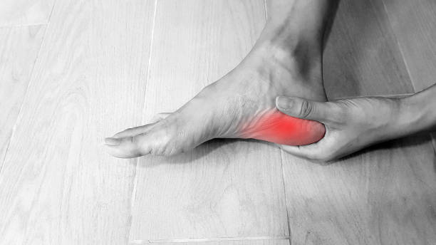

A plantar fasciitis doctor looks beyond the painful spot and studies how the entire foot handles weight. Several physical and imaging tests help confirm plantar fasciitis while ruling out conditions that require a different treatment plan.
Palpation of the medial heel to pinpoint localized tenderness
Gentle pressure along the inside and bottom of the heel helps locate the exact source of discomfort. Plantar fasciitis commonly produces tenderness near the medial calcaneal tubercle, where the plantar fascia attaches to the heel bone. Pain that appears in the center of the heel, along its outer border, or farther into the arch may suggest another condition.
Careful palpation also shows whether symptoms remain confined to one point or spread along a nerve or tendon. Swelling, warmth, tissue thickening, and sensitivity on both sides of the heel provide additional clues. An Athens podiatrist compares these findings with the patient’s morning pain, work demands, exercise habits, and recent changes in activity.
Windlass test to reproduce fascia pain through big toe extension
During the Windlass test, the examiner gently lifts the big toe while the patient sits or stands. That motion tightens the plantar fascia and raises the arch, reproducing the same mechanical tension created during push-off. Familiar pain near the heel attachment supports the diagnosis, particularly when symptoms match the usual first-step pattern.
Standing makes the test more demanding because body weight adds pressure through the foot. Limited toe movement, arthritis, or guarding can affect the result, so a negative response does not always rule out plantar fasciitis. A plantar fasciitis specialist interprets the test alongside other examination findings rather than treating it as proof by itself.
Dynamic gait analysis to evaluate pronation and foot mechanics
Walking analysis reveals how the heel, arch, ankle, and toes move through each phase of a step. Excessive pronation may stretch the plantar fascia, while a rigid high arch can reduce shock absorption and concentrate force beneath the heel. Uneven stride length or early heel lift may expose restrictions that remain hidden during a seated examination.
Video recordings, pressure measurements, and visual observation can all support a gait assessment. Shoe wear patterns may add useful context by showing repeated loading along one edge or beneath a specific area. A podiatrist in Athens AL uses this information to decide whether footwear changes, orthotics, strengthening, or mobility work could reduce recurring strain.
Diagnostic ultrasound to measure fascia thickness and swelling
High-frequency ultrasound creates a detailed image of the plantar fascia without radiation. Thickening near the heel, disrupted fibers, surrounding fluid, and changes in tissue texture may support a diagnosis of chronic plantar fasciitis. Comparison with the opposite foot can help determine whether the measurement represents a meaningful abnormality.
Real-time imaging also allows the clinician to watch the tissue while the toes and ankle move. Small tears, calcified deposits, or nearby soft-tissue problems may become visible during the scan. Because ultrasound findings do not always match pain severity, the examination and symptom history remain essential.
Weight-bearing X-rays to check for bone spurs or stress fractures
Standing X-rays show how the bones and joints align under normal body weight. Images may reveal heel spurs, arthritis, arch collapse, unusual bone position, or signs of an older injury. Although standard radiographs do not display the plantar fascia clearly, they help exclude structural causes of heel pain.
Early stress fractures may remain invisible until healing changes develop. Persistent tenderness around the sides of the heel can therefore lead a plantar fasciitis doctor to order follow-up imaging even after a normal X-ray. Heel spurs also require careful interpretation because many painless feet have them.
Foot and ankle MRI to detect deep soft tissue tears or inflammation
MRI provides detailed views of the fascia, tendons, muscles, nerves, heel bone, and surrounding tissue. A scan may show plantar fascia thickening, partial tearing, deep inflammation, bone marrow swelling, or muscle changes linked to nerve compression. This level of detail becomes valuable when symptoms remain severe or fail to respond to appropriate care.
Cost and testing time usually prevent MRI from serving as the first examination for routine heel pain. Persistent nighttime discomfort, unexplained weakness, trauma, or an unusual pain pattern may justify the added imaging. Results help separate plantar fasciitis from stress injury, tendon disease, tumors, and Baxter nerve entrapment.
Ankle dorsiflexion test to assess calf and Achilles tightness
Dorsiflexion testing measures how far the lower leg can move forward over the foot while the heel stays down. Restricted motion often points to tight calf muscles, a shortened Achilles tendon, or stiffness within the ankle joint. Each limitation can force the foot to compensate and place added tension across the plantar fascia.
Separate testing with the knee straight and bent helps identify which calf structure restricts movement. Weight-bearing assessment may provide a more realistic picture of what happens during walking than a seated measurement alone. The Foot Clinic can combine hands-on testing, gait evaluation, imaging, and mobility checks to determine whether plantar fasciitis or another heel condition is responsible for a patient’s symptoms.